Side Effect Registry Immuno-Oncology

Side Effect Registry Immuno-Oncology

SERIO is a registry for adverse events of immunotherapies (immune-related adverse events, irAE).

In particular, we aim to collect cases of rare, complex and therapy-refractory adverse events as well as adverse events in special patient groups (i.e. patients with autoimmune diseases or patients with a solid organ transplant).  Immunotherapies include checkpoint-inhibitors, cellular therapies, e.g. CAR T cells, and bispecific antibodies.

Our goal is to improve side effect management, gain insights into the pathogenesis of irAE, and be able to make predictions about irAE.
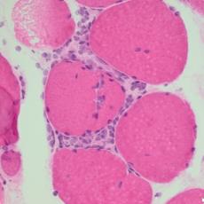

Myositis
Arthritis
Arthritis
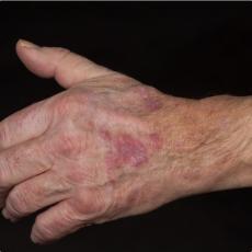
Lichen ruber
Lichen ruber
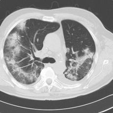
Pneumonitis
Pneumonitis

Who we are

SERIO is based at the Department of Dermatooncology at the University Hospital of Munich (LMU). The SERIO online register is operated in cooperation with the Paul Ehrlich Institute. Over the past 13 years, we have collected more than 1832 cases of rare, complex or very severe side effects from 27 centres in 6 countries. We cooperate with side effect specialists from all over the world.

SERIO was first initiated in 2011 by dermatooncologists from the University Hospital of Erlangen. The first documented case of SERIO dates back to 2009. A close collaboration with endocrinologists, cardiologists and gastroenterologists soon developed, which led to the initiation of our interdisciplinary Tox Board. Our cooperation with the Working Group Dermatooncolgy (ADO) includes the implementation of joint projects and the exchange of experiences.

SERIO aims to help physicians manage side effects and gain better knowledge of side effects induced by immunotherapy.

What we do 

  • Collect and analyze cases of rare, complex, severe and therapy-refractory adverse reactions induced by immunotherapy
  • Provide physicians with recommendations for the management of irAE
  • Conduct research and assist others conducting research related to irAE


          1533 - irAEs            73 - centers                         17 - tumor types

Case of the Month

60-year-old male patient with Nivolumab-induced polyarthritis and suspected necrotizing fasciitis

Medical history

Metastatic cutaneous melanoma, stage IV, M1d (AJCC 2017), BRAF V660K/V600R/V600M mutated; metastases in the brain, right parotid, lymph nodes and soft tissue

ECOG 0

Oncological Therapy

2006 Resection and lymph node resection

05/2019 Right parotid metastasis (right neck dissection)

07/2019-07/2020 Pembrolizumab adjuvant

08/2021 Recurrence (lymph node and soft tissue metastases)

08/2021-11/2021 Ipilimumab/nivolumab

11/2021-03/2022 Nivolumab, discontinuation due to recurrence (soft tissue metastasis)

05/2022-08/2023 Dabrafenib/trametinib

02/2023-08/2023 Stable disease

08/2023 Brain metastasis right temporal

09/2023-10/2023 Two cycles dacarbazine (DTIC)

10/2023 Reinduction of immunotherapy with ipilimumab/nivolumab as well as stereotactic irradiation of single cerebral metastases

12/2023 Diarrhea (approx. 10 stools/day), ICI-induced, improvement after a short prednisolone course with 1 mg/kg body weight

01/2024-04/2024 Nivolumab 240mg every 2 weeks

05/2024 Double dose of nivolumab (480mg every 4 weeks)

Immune-related adverse event

Approximately 1.5 weeks after receiving the double dose of nivolumab, the patient developed intermittent large joint pain (shoulders, knees, hips) and severe swelling and overheating of the right knee joint and the adjacent dorsal lower leg. Low dose cortisone (20 mg prednisolone/day) only led to a temporary, slight improvement in symptoms. He was admitted to a regional hospital. CT showed multiple air pockets on the medial head of the gastrocnemius muscle and several knee punctures excluded infectious cause. Due to the detected intraarticular air, severe pain in the right calf, swelling and overheating of the area around the right knee and reduced general condition, necrotizing fasciitis was suspected, so soft tissue surgery was performed on the right lower leg. The surgeons found an old hematoma, secretion, glassy disintegration of the m. sartorius, m. soleus and the m. gastrocnemius fascia, and an initial muscular disintegration of the medial m. gastrocnemius head. Histologically, there was no evidence of malignancy and there was no proof of streptococcus pyogenes or other bacteria. After the surgery, the patient was monitored in the intensive care unit and received clindamycin and piperacillin/tazobactam i.v. because the inflammatory parameters were elevated.

How we proceeded

After discharge the patient presented to our oncology outpatient clinic in a wheel chair with severe pain of the knee joints, weakness and pain in the muscles, especially the shoulders and the right calf. Joints were swollen with minimal effusions. Upon induction of prednisolone (1mg/kg body weight) the pain stopped within one day and the patient could get up from the wheel chair again. The initial radiological findings were re-evaluated and synovitis was diagnosed, without evidence of necrotizing fasciitis. The effusion in the knee showed 6,200 leukocytes/µl and an aviscous consistency, without crystals or bacteria. In summary, the patient suffered from myositis and polyarthritis induced by the checkpoint inhibitor therapy and subsequently an iatrogenic infection in the area of ​​the arthroscopic joint puncture. Prednisolone/d was tapered and methotrexate 15mg/week s.c. initiated. If this therapy does not significantly improve symptoms or the glucocorticoids cannot be tapered off, therapy with tocilizumb will be considered.

Could we continue the therapy?

Since the patient is still suffering from the adverse event at the time of reporting and currently receives prednisolone in combination with methotrexate, the immune checkpoint inhibitor therapy is currently paused. Depending on the clinical findings, however, reinduction will be discussed later due to the advanced tumor state of the melanoma.

Background

Rheumatic and musculoskeletal immune related adverse events (irAEs) are observed in about 10% of patients receiving immune checkpoint inhibitors (ICIs) [1]. Musculoskeletal irAEs show a large clinical spectrum [2], are often steroid-refractory [3] and frequently persist after cessation of ICI therapy [4]. Myositis is important to monitor since it can occur concomitantly with cardiomyositis[2].

In case of steroid-refractory rheumatic irAE several conventional synthetic disease-modifying antirheumatic drugs (csDMARD) have been used and so far, no specific drug has proven superiority. Methotrexate was the most frequently drug prescribed, followed by hydroxychloroquine then sulfasalazine, either as monotherapy or in combination [1]. No safety issues were described regarding long-term use of methotrexate associated with ICI in a few patients, with a median follow-up of over 1 year [5]. However, it takes around 4-8 weeks for the full effect to occur, so patients often have to continue taking a steroid in a tapering dosage during this period [1].

In severe rheumatic and systemic irAE or with insufficient response to csDMARD, biological disease-modifying antirheumatic drugs (bDMARD) may be considered, with TNF or IL-6 inhibitors being the preferred options for inflammatory arthritis[6]. However, prolonged use of TNF inhibitors can induce widespread, significant immunosuppression, which can negatively impact the antitumor efficacy of ICI therapy [7]. The anti-interleukin (IL)-6 receptor antibody, tocilizumab, is approved for the treatment of rheumatoid arthritis and juvenile idiopathic arthritis. It is reported to drastically improve symptoms for patients with severe ICI-induced polyarthritis [7].

The decision to reinduce the ICI-therapy should be based on the severity of musculoskeletal irAE, the extent of required immunosuppressive regimen and response of the irAE, the tumor dynamics, as well as therapy alternatives[1]. A pre-existing autoimmune rheumatic and/or systemic disease should not preclude the use of cancer immunotherapy. Baseline immunosuppressive regimen should be kept at the lowest dose possible (for glucocorticoids, below 10 mg prednisone per day) [1].

Our conclusion

This patient was thought to have a severe infection leading to necrotizing fasciitis, whereas his muskuloskeletal irAE quickly improved upon initiation of corticosteroids. Great care is needed before initiating immunosuppression in suspected infection. Thus, clarifying the genesis between an infectious and an autoimmune cause of the symptoms during ICI therapy is essential.

References

1.            Kostine, M., et al., EULAR points to consider for the diagnosis and management of rheumatic immune-related adverse events due to cancer immunotherapy with checkpoint inhibitors. Ann Rheum Dis, 2021. 80(1): p. 36-48.

2.            Moreira, A., et al., Myositis and neuromuscular side-effects induced by immune checkpoint inhibitors. Eur J Cancer, 2019. 106: p. 12-23.

3.            Tomsitz, D., et al., Steroid-Refractory Immune-Related Adverse Events Induced by Checkpoint Inhibitors. Cancers (Basel), 2023. 15(9).

4.            Schulz, T.U., et al., Persistent immune-related adverse events after cessation of checkpoint inhibitor therapy: Prevalence and impact on patients' health-related quality of life. Eur J Cancer, 2022. 176: p. 88-99.

5.            Leipe, J., et al., Characteristics and treatment of new-onset arthritis after checkpoint inhibitor therapy. RMD Open, 2018. 4(2): p. e000714.

6.            Grumme, L. and H. Schulze-Koops, [Rheumatological side effects of checkpoint inhibitors and their treatment]. Z Rheumatol, 2023. 82(3): p. 187-194.

7.            Kim, S.T., et al., Successful treatment of arthritis induced by checkpoint inhibitors with tocilizumab: a case series. Ann Rheum Dis, 2017. 76(12): p. 2061-2064.

44-year old female patient with pembrolizumab-induced polyserositis

Medical history

10/21 superficial spreading melanoma (Breslow thickness: 1.5mm), localization: upper back

Axillary Sentinel Lymph Node Biopsy left (1/1) and right (0/1), pT2a N1a M0, St. IIIB (AJCC 2017)

BRAF V600E

Oncological Therapy

12/2021 – 09/2022 – adjuvant therapy with pembrolizumab (12 cycles) 

Immune-related adverse event

11 months after initiation of immunotherapy with pembrolizumab, the patient presented with pleural effusion and ascites. First, steroid therapy was initiated. During steroid tapering, the patient developed recurrent chylous pleural effusion and ascites. Steroid therapy was re-initiated but without success. Spironolactone, antibiotics, and a fat-free diet were initiated. Steroid therapy was stopped. Subsequently, the patient presented with severe dyspnea and fever due to recurrent chylous pleural effusion and ascites. Repeatedly pleural effusions and ascites were drained yielding up to 5 liter of fluids with no evidence for malignant cells. Staging and assessment of the tumor marker S100 revealed no progression of disease. The young patient had to quit work and has been in and out of hospitals for 5 months. She also appeared cachectic upon our first consultation.

How we proceeded

Since symptoms were steroid-refractory upon relapsing symptoms during steroid taper and the second increase of steroids we initiated systemic therapy intravenous immunoglobulins (IVIG).  After 4 cycles, we started parallel treatment with Extracorporeal Photopheresis (ECP). Lymphangiography was planned to rule out lymphatic fistulae. The patients` condition has significantly improved upon start of IVIG in combination with ECP. She currently reports no dyspnea, with less ascites.  She adheres to a low-fat, high-protein diet. ECP and IVIG therapies are ongoing.

Therapy with ruxolitinib could be considered in the future. Follow-up nutritional and metabolic consultations are scheduled to evaluate the further approach regarding the fat-free diet.

Could we continue the immunotherapy?

The checkpoint inhibitor therapy was permanently discontinued.

Background

The patient presents with a complex case of checkpoint-inhibitor induced polyserositis with recurrent chylous pleural effusions and ascites induced by adjuvant anti-PD1 therapy. The treatment approach has involved a combination of immunomodulatory and nutritional interventions in addition to repeated drainage of the effusions.

IrSerositis is a rare side effect of immunotherapy and can induce potentially life-threatening side effects with cardiac and pulmonary affection (Zierold et al., 2022). Knowledge about this immune-related adverse event is scarce. It can manifest acutely or progress slowly, occurring between 7 days and up to 72 or even 200 weeks after initiation of immunotherapy (Zierold et al., 2022). Concurrent malignant infiltrates are possible, emphasizing the importance of histopathological analysis (presence of lymphocytes, absence of malignant cells) (Zierold et al., 2022). Treatment of irSerositis can be challenging. High-dose steroid therapy could be a viable primary treatment option. Nevertheless, frequent pleural or ascites drainages are often required. For steroid-refractory immune-related adverse events (irAEs), a second-line therapy may be initiated (e.g., Infliximab, Ruxolitinib). Once, spontaneous resolution was reported (Kolla et Patel, 2016). Tumor outcomes during treatment of irSerositis vary (Zierold et al., 2022).

Our conclusion

Polyserositis is a very rare side effect of immune checkpoint inhibitors. Prompt diagnosis with exclusion of malignant cause and treatment are essential. The patient's positive response to the current treatment regimen with IVIG and ECP is encouraging.

56-year old melanoma patient with positive test for purkinje cell cytoplasmatic antibody

Medical history

04/22 nodular melanoma

pT4b pN3c M1b, St. IV (AJCC 2017)

BRAF mutated

Oncological Therapy

04/2022 tumor resection (R1) and axillary lymph node dissection

06-08/2022 monotherapy with nivolumab

06/2022 tumor resection (R0)

08-09/2022 radiotherapy

08-09/2022 2 cycles of combined immunotherapy with ipilimumab 3mg/kg + nivolumab 1mg/kg (stopped)

Since 12/2022 BRAF/ MEK inhibitor therapy with BRAFTOFI (Encorafenib) + MEKTOVI (Binimetinib)

Immune-related adverse event

After 2 doses of combined immunotherapy with ipilimumab and nivolumab, the patient presented with immune-related hepatitis (irHepatitis), immune-related hypophysitis (irHypophysitis) and immune-related myocarditis (irMyocarditis). Subsequently prednisolone treatment was initiated. Due to the high dose corticosteroid therapy, the patient developed steroid-related diabetes mellitus.

In further laboratory tests to exclude other conditions, we report a newly discovered serum reactivity to purkinje cell cytoplasmic antibodies without anti-Yo specifity. Control analyses were performed to rule out a panel of previously described paraneoplastic antibodies known to be associated with paraneoplastic cerebellar degeneration. The patient presented without any neurological symptoms. Cranial MRI was normal.

As corticosteroid therapy improved symptoms of irMyocarditis and irHepatitis, prednisolone was tapered. For irHypophysitis treatment, hormone replacement therapy was continued with hydrocortisone. BRAF/ MEK inhibitor therapy was initiated.

How we proceeded

We initiated a systemic therapy with prednisolone with an initial dose of 80mg/day and then tapered.

Hypophysitis treatment usually involves lifelong hormone replacement therapy. Hydrocortisone with a dose of 30mg/day was substituted. Due to the risk of acute adrenal crisis, patients needs to be educated to increase the dose in stress situations (infections, trauma etc.) and should carry an emergency card with them at all times.

If a patient has a positive test for purkinje cell cytoplasmic antibodies, a follow-up with FDG-PET/CT to identify any underlying lesion is advised. Further neurological assessments are planned.

Could we continue the immunotherapy?

The immunotherapy was permanently discontinued due to the risk of irMyocarditis, irHypophysitis and irHepatitis. Positive tests for purkinje cell cytoplasmic antibodies often appear to be unrelated to the development of neurological diseases, therefore interruption of immunotherapy may not be required.

Background

Paraneoplastic cerebellar degeneration is an uncommon disorder that can be associated with different types of cancer. The most commonly associated cancers are small cell lung cancer, gynecologic cancer and lymphoma. Purkinje cell cytoplasmic antibody type 1 with anti-Yo specifity (PCA-1) predominantly associate with cerebellar degeneration (Wolters Kluwer et al., Uptodate, 2023).

But, a positive test for paraneoplastic antibodies, as the purkinje cell cytoplasmic antibody, does not indicate the presence or development of a neurological disease. Many patients can have a positive test at low titers.  Even when detected at high titer, neurological symptoms or physical findings are not mandatory (Wolters Kluwer et al., Uptodate, 2023). To identify first signs of a neurological disorder, regular physical examinations are advised. FDG-PET/CT should be performed once to rule out cerebellar degeneration. MRI scans are often unremarkable.

Case reports reported of ipilimumab induced cerebellar degeneration in lung cancer patients (Hardwick et al., Neuropathology and Applied Neurobiology, 2021). Purkinje cell cytoplasmic antibody type 1 with anti-Yo specifity (PCA-1) tests were detected at low titer. Symptoms included severe ataxia, dysarthria, dizziness and double vision. Methylprednisolone and infliximab were initiated to improve symptoms.

Generally, treating the underlying cancer is the best way to control a paraneoplastic cerebellar degeneration (Elsevier, ScienceDirect, 2023).

Our conclusion

A positive test for paraneoplastic antibodies is a very rare side effect of immune checkpoint inhibitors. Further research is now needed to evaluate the exact role of purkinje cell cytoplasmic antibodies without anti-Yo specifity. Neurological monitoring has to be indicated to detect changes in the clinical status of tumor patients.

56-year old male patient with ipilimumab/ nivolumab-induced adrenalitis

Medical history

05/22 melanoma of unknown primary (MUP)

Manifestation: Lymph node metastasis right axilla

TxN3M1b (1), (AJCC 2017)

Staging cMRT and CT-thorax/abdomen: Pulmonary lymph node metastases

Oncological Therapy

06-08/22 – 4 cycles of combined immunotherapy with ipilimumab 3mg/kg + nivolumab 1mg/kg

Since 08/22 – monotherapy with nivolumab (paused 11-12/22)

Immune-related adverse event

10 weeks after initiation of combined immunotherapy with ipilimumab and nivolumab, the patient presented with nausea, fatigue and impaired vision. First, immune-related hypophysitis was suspected (irHypophysitis) and subsequently hydrocortisone treatment was initiated. Later on the patient developed increasing hyponatraemia and impaired renal function. The hydrocortisone therapy was continued.

The endocrinological examination showed low cortisol (0,5 µg/dl; reference range: 6,2-18,0 µg/dl), and increased adrenocorticotropic hormone (ACTH) levels (570 pg/ml; reference range: 4-61 pg/ml)). As the ACTH provocation test was negative (low cortisol (0,6 µg/dl) 30min after administration of ACTH, irHypophysitis was ruled out and irAdrenalitis was diagnosed with primary adrenocortical insufficiency.

As hydrocortisone therapy improved symptoms, nivolumab was re-initiated without any further complications. Sodium, glomerular filtration rate (GFR) and creatinine returned to levels within the normal range.

How we proceeded

We initiated systemic therapy with hydrocortisone with a dose of 30mg/day (20-10-0mg, later 20-5-5mg). Importantly, individual dose adjustments of hydrocortisone may be necessary (e.g. illness, infection, operation, stress, hyperthyreodism, etc.). Mineralocorticoids were replaced by fludrocortisone (Astotin H 0,05mg/day). Based on individual symptoms like decreased well-being or low libido, some patients also take dehydroepiandrosterone.

Could we continue the immunotherapy?

Nivolumab monotherapy was temporarily paused until symptoms subsided. In combination with the administration of hydrocortisone and fludrocortisone, Nivolumab was re-initiated without complications.

Background

Endocrine adverse events occur in approximately 10% of patients treated with combined immunotherapy (Hattersley et al., ClinMed, 2021), thyroiditis (10-16%) and hypophysitis (17%) being the most common (Husebye et al., European Journal of Endocrinology, 2023). Adrenalitis with primary adrenal insufficiency (1%), diabetes mellitus and diabetes insipidus (0,9-2%) are rare however critical to diagnose (Husebye et al., European Journal of Endocrinology, 2022). Most endocrinopathies present with non-specific symptoms like fatigue, nausea, headache and arthralgia and can thus be missed (Braun GS et al., Nephrologe, 2020; Heinzerling et al., Dtsch Arztebl Int, 2019; Hattersley et al., ClinMed, 2021). Immunotherapy may be paused temporarily during endocrinological assessments.

Decreased fasting cortisol (morning) < 5 μg/dl and a change of less than 9 µg/dl after administration of 250µg of ACTH provide diagnostic clues for primary adrenal insufficiency (PAI) caused by i.e. irAdrenalitis. The classic short corticotropin test with ACTH is considered as the gold standard method to confirm the PAI. The electrolytes should also be checked as hyponatraemia and hyperpotassaemia can occur.

In the differential diagnosis of adrenal insufficiency, irHypophysitis has to be considered initially. IrHypophysitis occur in approximately 17% of patients receiving combined ICI therapy. If hypophysitis is suspected, checks for clinical signs like new severe headaches or complaints of vision changes, biochemical assessment of the hypothalamic-pituitary axis and MRI scan of pituitary are recommended (Husebye et al., European Journal of Endocrinology, 2022). Neurological examination should be performed once to rule out any other neurological causes. The pituitary hormones (GH, prolactin, LH, FSH, TSH, and ACTH) as well as target hormones should be measured. The corticotropic axis is most commonly affected. Low fasting cortisol (morning) 7-16 µg/dl and serum ACTH value not higher than normal (reference range 4-61 pg/ml) establish the diagnosis of secondary adrenal insufficiency caused by i.e. hypophysitis (Hattersley et al., ClinMed, 2021, Wolters Kluwer et al., UpToDate, 2023).  

Treatment usually involves lifelong hormone replacement therapy (corticosteroid and mineralocorticoid). Due to the risk of acute adrenal crisis, patients will need to be educated to increase the dose in stress situations (infections, trauma etc.) and should carry an emergency card with them at all times.

Our conclusion

Adrenalitis is a rare side effect of immune checkpoint inhibitors. Prompt diagnosis and treatment with hydrocortisone as well as patient education is essential.